| Back |
The ClearPET/XPAD project:
|
While X-ray computerized tomography (CT) allows imaging the mass density of living tissues using an external X-ray tube, positron emission tomography (PET) is imaging gamma rays directly emitted from the tissues. In this case, the emission of gamma rays results from the decay of radioactive nuclei used to label a radiopharmaceutical injected to the patient. Thus, contrary to transmission X-ray tomography, emission tomography images the function rather than the structure of living tissues. The development of combined PET/CT imaging systems led to a rapid expansion of this technique in clinical routine. Similarly, the development of dedicated PET scanners and micro-CT scanners for small animals pleads for joining these two modalities in a common gantry. |
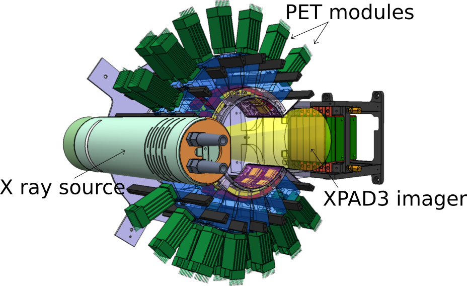 Layout of the ClearPET/XPAD prototype |
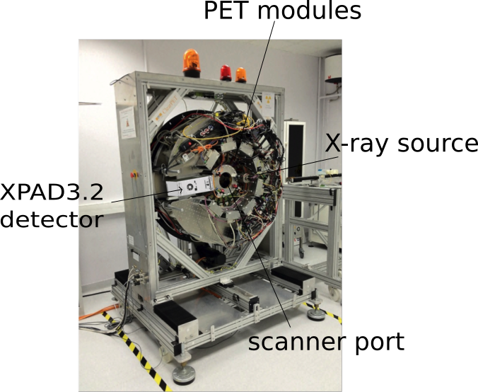 Picture of the ClearPET/XPAD prototype |
However, like with clinical PET/CT, the juxtaposition of both modalities does not allow for extrapolating the exact position of the animal during the PET scan. Therefore, we have combined the detector module of the ClearPET prototype scanner developed within the Crystal Clear Collaboration with the XPAD3 X-ray hybrid pixel detector in a common rotating gantry. To achieve this goal, we redesigned the partial ring arrangement of the ClearPET detectors. As a result, both detection systems have been merged together with an X-ray tube in a fully integrated PET/CT device that makes it possible to acquire simultaneous emission and transmission scans for mice. |
As an example, a simlultaneous PET/CT scan of a live mouse was performed after a 100 μL injection of 18 MBq [18F]FDG in the tail vein. Following a 45 minutes uptake phase, images were acquired during 60 minutes with the mouse under isofluorane anesthesia. The Mo anode X-ray tube was operated at 50 kV and 20 μA with a filter of 500 μm aluminum. The reconstructed transverse and sagittal slices through the mouse at the heart level show FDG uptake in the myocardium and brown fat, as well as the left Harderian gland (sagittal slice). |
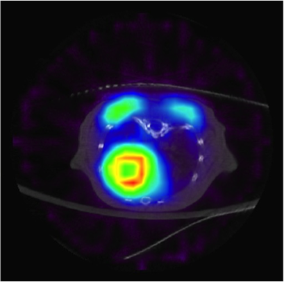 Transverse slice |
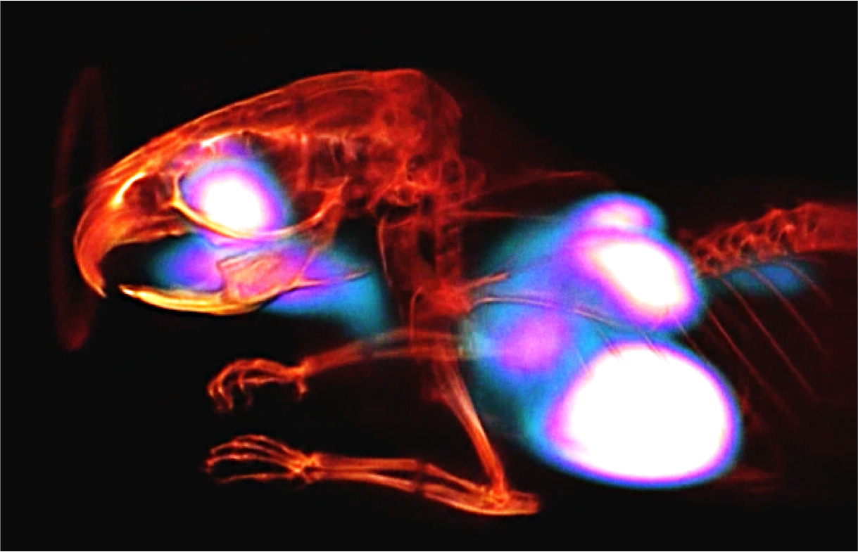 3D rendering |
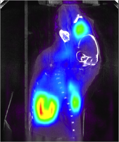 Sagittal slice |
Master Thesis
Jérôme Loess, Mise en œuvre et carctérisation de la caméra TEP pour petits animaux ClearPET, Université de la Méditerranée Aix-Marseille II, juin 2006
Eric Guedj, Normalisation géomérique et en efficacité des images obtenues par le ClearPET/XPAD, système d'imagerie hybride pour petits animaux, permettant une acquisition TEP/TDM simultanée, Ecole Supérieure d'Ingénieurs de Luminy (ESIL), septembre 2010
Margaux Hamonet, Normalisation et correction d'atténuation lors de l'acquisition TEP/TDM simultanée avec le prototype ClearPET/XPAD, Université de Nantes, août 2012
Isabell Bones, Performance evaluation of the prototype scanner ClearPET/XPAD for simultaneous PET/CT imaging, Erasmus+ internship, Fach Hochschuhle Aachen, Apr 2016
Borana Mom, Simulation Monte Carlo d'un TEP basé sur l'annihilation en trois photons, Université Claude-Bernard Lyon I, septembre 2019
PhD Thesis
Jean-Baptiste Mosset, Développement d'un module de détection phoswich LSO/LuYAP pour le prototype de caméra à positrons ClearPET, Ecole Polytechnique Fédérale de Lausanne (EPFL), juillet 2006
Martin Rey, Etude du tomographe de haute résolution pour petits animaux ClearPET par la méthode de Monte Carlo, Ecole Polytechnique Fédérale de Lausanne (EPFL), juin 2007
Stan Nicol, Etude et construction d'un tomographe TEP/TDM pour petits animaux, combinant modules phoswich à scintillateurs et détecteur à pixels hybrides, Université de la Méditerranée Aix-Marseille II, juillet 2010
Margaux Hamonet, Tomographie hybride simultanée TEP/TDM combinantdétecteurs à pixels hybrides et modules phoswich à scintillateurs, Aix-Marseille Université, avril 2016
Conference Records
E. Auffray et al., The ClearPET project, in Conf. Rec. ITBS 2003, 26-30 May, Milos Island, Greece, Nucl. Instr. Meth. Phys. Res. A 527 (2004) 180-189
S. Weber et al., Image reconstruction for the ClearPET Neuro, in Conf. Rec. ITBS 2005, 25-29 Sep, Milos Island, Greece, Nucl. Instr. Meth. Phys. Res. A 569 (2006) 381-385
D. Wisniewski et al., Digital pulse shape discrimination methods for phoswich detectors, in IEEE NSS/MIC 2005, 23-29 Oct, San Juan, Puerto Rico Island, IEEE Press, pp. 2979-2983
M. Rey et al., Measured and simulated specifications of the Lausanne ClearPET scanner demonstrator, in IEEE NSS/MIC 2005, 23-29 Oct, San Juan, Puerto Rico Island, IEEE Press, pp. 2070-2073
M. Khodaverdi et al., High resolution imaging with ClearPETTM Neuro - First animal images, in IEEE NSS/MIC 2005, 23-29 Oct, San Juan, Puerto Rico Island, IEEE Press, pp. 1641-1644
M. Rey et al., Count rate performance study of the Lausanne ClearPET scanner demonstrator, in Conf. Rec. EuroMedIm 2006, 9-12 May, Marseille, France, Nucl. Instr. Meth. Phys. Res. A 571 (2007) 207-210
M. Khodaverdi et al., Design study for the,ClearPET/XPAD small animal PET/CT scanner, in IEEE NSS/MIC 2007, 27 Oct-3 Nov, Honolulu, Hawaii, IEEE Press, pp. 4300-4302
S. Nicol, S. Karkar, P. Descourt, C. Morel, The ClearPET/XPAD: development of a simultaneous PET/CT scanner for mice, in Conf. Rec. ESMI TOPIM 2009, 26-30 Jan, Les Houches, France, p. 29
S. Nicol et al., Design and construction of the ClearPET/XPAD small animal PET/CT scanner, in IEEE NSS/MIC 2009, 25-31 Oct, Orlando, Florida, IEEE Press, pp. 3311-3314
M. Hamonet et al., The ClearPET/XPAD prototype: development of a simultaneous PET/CT scanner for mice, in Conf. Rec. IEEE NSS/MIC 2015, 31 Oct-7 Nov, San Diego, California, USA, IEEE Press
Publications
K. Ziemons et al., The ClearPET project: development of a 2nd generation high- performance small animal PET scanner, Nucl. Instr. Meth. Phys. Res. A 537 (2005) 307-311
J.-B. Mosset et al., Development of an Optimized LSO/LuYAP Phoswich Detector Head for the Lausanne ClearPET Demonstrator, IEEE Trans. Nucl. Sci. 53 (2006) 25-29